BMC Pulmonary Medicine
Danilo Buonsenso,
Anna Maria Musolino,
Antonio Gatto,
Ilaria Lazzareschi,
Antonietta Curatola &
Piero Valentini
Abstract
Lung ultrasound (LUS) is nowadays a fast-growing field of study since the technique has been widely acknowledged as a cost-effective, radiation free, and ready available alternative to standard X-ray imaging. However, despite extensive acoustic characterization studies and documented medical evidences, a lot is still unknown about how ultrasounds interact with lung tissue. One of the most discussed lung artifacts are the B-lines [in all ages] and the subpleural consolidations (in young infants). Recently, LUS has been claimed to be able to detect pneumonia in infants with bronchiolitis, although this can be an overestimation due to the peculiar physiology of small peripheral airways of the pediatric lung (particularly in neonate/infants).
 |
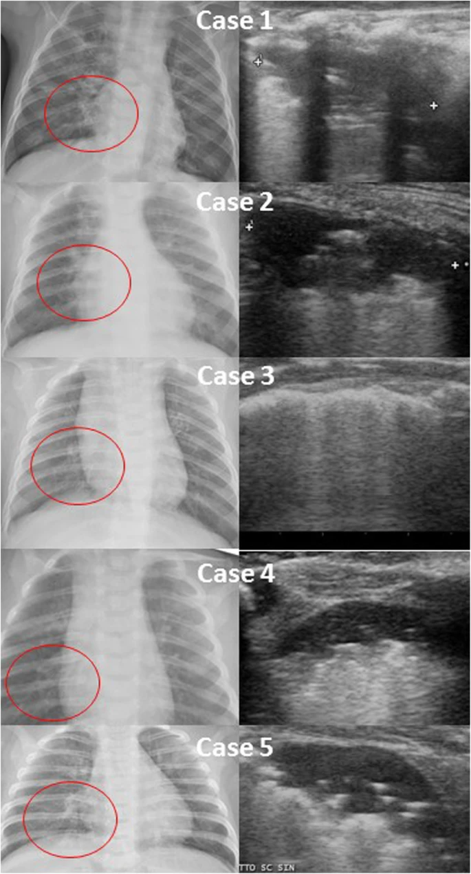
| A case series of paracardiac consolidations on chest X-ray and the corresponding pattern on lung ultrasound, showing posterior, paravertebral, subpleural consolidations |
No comments:
Post a Comment