Background
Asthma is a common chronic airway disease in the world. The purpose of this study was to explore the expression of IL1-RL1 in sputum and its correlation with Th1 and Th2 cytokines in asthma.
Methods
We recruited 132 subjects, detected IL1-RL1 protein level in sputum supernatant by ELISA, and analyzed the correlation between the expression level of IL1-RL1 and fraction of exhaled nitric oxide (FeNO), IgE, peripheral blood eosinophil count (EOS#), and Th2 cytokines (IL-4, IL-5, IL-10, IL-13, IL-33 and TSLP) and Th1 cytokines (IFN-γ, IL-2, IL-8). The diagnostic value of IL1-RL1 was evaluated by ROC curve. The expression of IL1-RL1 was further confirmed by BEAS-2B cell in vitro.
Results
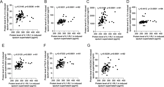
Compared with the healthy control group, the expression of IL1-RL1 in sputum supernatant, sputum cells and serum of patients with asthma increased. The AUC of ROC curve of IL1-RL1 in sputum supernatant and serum were 0.6840 (p = 0.0034), and 0.7009 (p = 0.0233), respectively. IL1-RL1 was positively correlated with FeNO, IgE, EOS#, Th2 cytokines (IL-4, IL-5, IL-10, IL-13, IL-33 and TSLP) and Th1 cytokines (IFN-γ, IL-2, IL-8) in induced sputum supernatant. Four weeks after inhaled glucocorticoids (ICS) treatment, the expression of IL1-RL1 in sputum supernatant and serum was increased. In vitro, the expression of IL1-RL1 in BEAS-2B was increased after stimulated by IL-4 or IL-13 for 24 h.
Conclusion
The expression of IL1-RL1 in sputum supernatant, sputum cells and serum of patients with asthma was increased, and was positively correlated with some inflammatory markers in patients with asthma. IL1-RL1 may be used as a potential biomarker for the diagnosis and treatment of asthma.


No comments:
Post a Comment