Abstract
Objective
To explore the role of different cells and molecules in the pathogenesis of allergic rhinitis (AR) with positive Artemisia allergen by detecting their expression levels.
Methods
From January 2021 to December 2022,200 AR patients diagnosed in the Otolaryngology Clinic of Ordos Central Hospital were selected as the AR group, and 50 healthy people who underwent physical examination in the hospital during the same period were randomly selected as the healthy control (HC) group. The levels of GATA-3mRNA, RORγtmRNA and FoxP3mRNA in peripheral blood mononuclear cells were detected by real-time fluorescence quantitative PCR (qRT-PCR). The proportions of Th2, Th17 and Treg cells were detected by flow cytometry. The concentrations of IL-4, IL-5, IL-17 and IL-10 in serum were detected by enzyme-linked immunosorbent assay.
The differences of transcription gene level, immune cell ratio and cytokine concentration between the two groups were analyzed.Results
 |
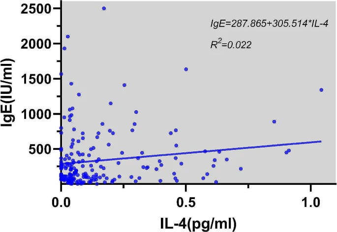
| Linear regression for the effect of IL-4 on IgE |
Conclusion
The secretion of immune cells and cytokines in peripheral blood of AR patients is abnormal. Th2, Th17, Treg specific transcription factors and related cells and cytokines are involved in the occurrence and development of allergic rhinitis.

No comments:
Post a Comment